
Hello Buddyz!

Since I was become a microbiology students at UITM KUALA PILAH,NEGERI SEMBILAN.. I became too busy and i have no time to do update my blog. Okay, back to the main topic,when i'm studying microbiology, many thing i've learnt and so many fun facts that i was dicovered. Thus, i want to share ONE of topic that you'll have fun when you've learnt.
WHAT IS MICROBIOLOGY?
 Microbiology is defined as "the study of organisms too small to be clearly seen by the under naked eye." These organisms include viruses, bacteria, algae, fungi, archaea, and protozoa. When studying microorganisms, microbiologists are concerned with characteristics and functions such as morphology, cytology, physiology, ecology, taxonomy, genetics, and molecular biology.


HONEY can CURE all type of illness including CANCER, IS IT TRUE?

Yes,it is true, THE HOLY QURAN prove it 14 centuries back!
and YOU will discover it when you've learnt:
MEDICAL MICROBIOLOGY AND IMMUNOLOGY
Islam emphasized on the importance of honey, the word (healing) appeared 4 times in Qur’an, 3 times linked to Qur’an and one time linked to honey. Allah the Almighty says “Then, eat of all fruits, and follow the says of your Lord made easy (for you). There comes forth from their bellies, a drink of varying colour wherein is healing for men. Verily, in this is indeed a sign for a people who think.” (An-Nahl:69).
and YOU will discover it when you've learnt:
I believed this and i'm know ALL TYPE OF CANCER CAN BE CURED by using HONEY, which anybody doesn't know.OKAY,LET ME PROVE ONE BY ONE:
Honey has power to fight cancer
Olivia Olarte-Ulherr / 25 February 2013
Scientific findings by researchers here could be the breakthrough the world has long been waiting for in cancer treatment.
 “Manuka honey has been recognised for its anti-bacterial and wound-healing properties for many years. However, the potential effect of manuka on cancer cells has not been investigated in detail,” said Dr Basel Al Ramadi, professor and chair of the Department of Medical Microbiology and Immunology, College of Medicine and Health Sciences at the UAEU.
Olivia Olarte-Ulherr / 25 February 2013
Scientific findings by researchers here could be the breakthrough the world has long been waiting for in cancer treatment.
The groundbreaking discovery by the research team at the UAE University found that honey from New Zealand’s manuka tree can effectively inhibit growth of cancer cells, including breast, skin and colon cancer; and tremendously reduce the toxicity associated with chemotherapy treatment.
“Manuka honey has been recognised for its anti-bacterial and wound-healing properties for many years. However, the potential effect of manuka on cancer cells has not been investigated in detail,” said Dr Basel Al Ramadi, professor and chair of the Department of Medical Microbiology and Immunology, College of Medicine and Health Sciences at the UAEU.
In the study, led by Dr Al Ramadi, the team of researchers used three different cancer cell lines (breast, skin and colon cancer) and demonstrated that the addition of exceedingly small amounts of manuka honey, as little as 1.0 per cent, can stop the growth of cancer cells by up to 70 per cent.
The team of investigators carried out further studies to characterise the mechanism by which manuka honey is inducing the death of cancer cells.
“The evidence so far suggests that manuka acts by stimulating a number of proteins inside the cells that leads to the induction of apoptosis, or programmed cell death. This is a natural process by which our body eliminates old or unwanted cells and is part of the normal organism’s development,” Dr Al Ramadi told Khaleej Times.
Using an experimental cancer model, in which mice are implanted with fast-growing skin tumour cells, the researchers administered manuka honey intravenously in conjunction with chemotherapy, and the results showed an improvement in the overall survival of the animal.
“It is significant that a honey can do this.... The manuka honey alone can inhibit cancer growth by 30 per cent, but when combined with chemotherapy, there was 61 per cent inhibition,” Dr Al Ramadi stressed.
The study, which was carried out over five years and published in the renowned scientific journal PLOS ONE early this month, is expected to stimulate further investigations on the use of manuka honey in cancer treament in humans.
“This is a very exciting area of research and we are optimistic about what these new developments may mean in terms of potential new treatments for certain types of cancer,” the professor said.
Moving forward, the research team hopes to get funding for their next course of investigations.
These include identifying the active components of the manuka honey that is inducing the growth-inhibiting effect, to understand the molecular pathways by which the honey is inducing the death of cancer cells, and the actual testing of manuka honey in humans through small-scale clinical trials.
The team aims to carry out the studies within the next two years.
But first, “we will be applying for further funding from the National Research Foundation and Terry Fox, and for ethical approval”, said Dr Al Ramadi.
This year’s Terry Fox Run, the annual cancer fundraising initiative, was held on Friday and generated Dh337,533. Some of the money raised was given to UAEU for research.
The UAEU research team is collaborating with colleagues in the departments of oncology and surgery in Tawam Hospital to continue their investigation. Tawam Hospital has the most comprehensive oncology treatment facility in the country, registering up to 80 per cent of the cancer cases in the UAE.
 THIS IS HONEY MOLECULE
Intravenous Administration of Manuka Honey Inhibits Tumor Growth and Improves Host Survival When Used in Combination with Chemotherapy in a Melanoma Mouse Model

 Initial experiments were done to study the physiochemical characteristics of the manuka honey. Different dilutions of manuka were prepared directly in tissue culture medium in which the B16.F1 melanoma cells are routinely cultured and tested for their pH and osmolarity. The studies revealed that manuka solutions of concentrations up to 5% (w/v) were physiological (data not shown). Thus, all subsequent in vitro studies were carried out using manuka concentrations in the range of 0.3% to 5%.
Initial experiments were done to study the physiochemical characteristics of the manuka honey. Different dilutions of manuka were prepared directly in tissue culture medium in which the B16.F1 melanoma cells are routinely cultured and tested for their pH and osmolarity. The studies revealed that manuka solutions of concentrations up to 5% (w/v) were physiological (data not shown). Thus, all subsequent in vitro studies were carried out using manuka concentrations in the range of 0.3% to 5%.Manuka inhibits growth of cancer cells
The potential effect of manuka on cancer cell proliferation was investigated using three tumor cell lines differing in type and origin, the murine melanoma (B16.F1) and colon carcinoma (CT26) cells and the human breast cancer (MCF-7) cell line. Cells were incubated with different concentrations of manuka (range 0.3 to 2.5%) for 24–72 hrs. As a positive control, cells were cultured with taxol at a final concentration of 10 or 50 ng/ml. As shown in Figure 1, the addition of as little as 0.3% manuka to cells in culture resulted in a significant decrease in the viability of B16.F1 cells (panels A–C). This inhibitory effect on cell viability was dependent on both manuka concentration and total incubation time. By as early as 24 hrs, the viabilities of B16.F1 cells cultured with manuka at final concentrations of 0.3, 0.6, 1.25 and 2.5% were 85%, 75%, 60% and 43% of control (no manuka) cultures, respectively (Fig. 1A). In contrast, over the same time period, the viabilities of B16.F1 cells cultured in presence of 10 ng/ml or 50 ng/ml of the antineoplastic drug taxol were reduced to 90% or 83% of control, respectively (Fig. 1A). The decreased cell viability was more pronounced as the time of culture increased to 48 hrs (Fig. 1B) or 72 hrs (Fig. 1C). At the latter time point, cell viability was reduced to 17% in cell cultures treated with 2.5% manuka (Fig. 1C). Under the same conditions, cells cultured with 10 ng/ml or 50 ng/ml taxol had a reduction in viability to 64% or 34% of control, respectively. Essentially similar results were also observed with the CT26 (panels D–E) and MCF-7 (panels F–G) cancer cell lines. These results demonstrate that in vitro treatment of cancer cells with low concentrations of manuka resulted in significant inhibition of cell proliferation.

 Download:
Figure 1. Inhibition of cancer cell proliferation by manuka honey.
B16.F1 (graphs A–C), CT26 (graphs D, E) and MCF-7 (graphs F, G) cells were plated at 5×10³ cells per well and incubated for 24 hr (graphs A, D, F), 48 hr (graph B) or 72 hr (graphs C, E, G) in the absence or presence of the indicated concentrations of manuka honey (range 0.3% to 5.0% w/v), or taxol (10 ng/ml or 50 ng/ml final concentration). At the end of the incubation period, cell viability was determined using CellTiter-Glo luminescent assay. Results are expressed as percentage viability in treated cell cultures compared to control, untreated, cells and are representative of 3 (for B16.F1 cells) or 2 (for CT26 and MCF-7 cells) independent experiments. Asterisks denote statistically significant differences in viability of experimental groups compared to control (*, p<0.05; **, p<0.01; ***, p<0.001).
doi:10.1371/journal.pone.0055993.g001
Next, experiments were undertaken to investigate the effect of co-treatment with manuka and taxol on proliferation of B16.F1 melanoma cells. Taxol (10 ng/ml) plus different concentrations of manuka (0.6% to 5.0%) were added simultaneously at the initiation of B16.F1 cell culture and cell viability was assessed 72 hrs later. As can be seen in Figure 2, increasing concentrations of manuka resulted in correspondingly lower cell viability, reaching a mean of 6% at the highest concentration of manuka used. Importantly, however, the extent of cell death achieved by a combination of taxol and manuka was essentially the same as seen with manuka alone (Fig. 2). This suggests that taxol and manuka act in an additive, not synergistic, manner when used in combination with melanoma cancer cells.
 Download:
Figure 2. Co-treatment with manuka and taxol results in additive effect.
B16.F1 cells were seeded at 1×10³ cells per well in a 96-well plate and incubated with the indicated concentrations of manuka, alone or in combination with taxol (10 ng/ml), for 72 hrs. Cell viability was determined using CellTiter-Glo luminescent assay. Results are expressed as percentage viability in treated cell cultures compared to untreated cells and are representative of 3 independent experiments. Asterisks denote statistically significant differences between corresponding cell cultures treated with each manuka concentration in absence or presence of taxol (*, p<0.05).
doi:10.1371/journal.pone.0055993.g002

Download:
Figure 1. Inhibition of cancer cell proliferation by manuka honey.
B16.F1 (graphs A–C), CT26 (graphs D, E) and MCF-7 (graphs F, G) cells were plated at 5×10³ cells per well and incubated for 24 hr (graphs A, D, F), 48 hr (graph B) or 72 hr (graphs C, E, G) in the absence or presence of the indicated concentrations of manuka honey (range 0.3% to 5.0% w/v), or taxol (10 ng/ml or 50 ng/ml final concentration). At the end of the incubation period, cell viability was determined using CellTiter-Glo luminescent assay. Results are expressed as percentage viability in treated cell cultures compared to control, untreated, cells and are representative of 3 (for B16.F1 cells) or 2 (for CT26 and MCF-7 cells) independent experiments. Asterisks denote statistically significant differences in viability of experimental groups compared to control (*, p<0.05; **, p<0.01; ***, p<0.001).
doi:10.1371/journal.pone.0055993.g001
Download:
Figure 2. Co-treatment with manuka and taxol results in additive effect.
B16.F1 cells were seeded at 1×10³ cells per well in a 96-well plate and incubated with the indicated concentrations of manuka, alone or in combination with taxol (10 ng/ml), for 72 hrs. Cell viability was determined using CellTiter-Glo luminescent assay. Results are expressed as percentage viability in treated cell cultures compared to untreated cells and are representative of 3 independent experiments. Asterisks denote statistically significant differences between corresponding cell cultures treated with each manuka concentration in absence or presence of taxol (*, p<0.05).
doi:10.1371/journal.pone.0055993.g002Manuka induces apoptosis in cancer cells
In the next series of experiments, we addressed the potential mechanism by which manuka was causing decreased cell viability. Loss of cell membrane asymmetry, detectable by Annexin V staining, represents one of the earliest events in apoptosis. B16.F1 cells were harvested at 24, 48, or 72 hrs after treatment with different concentrations of manuka honey (range 0.3% to 5.0%) or taxol (at a final concentration of 10 ng/ml), stained with Annexin V-FITC and PI, and subjected to flowcytometric analysis. As can be seen in Figure 3, there was a dose-dependent, and time-dependent, increase in the number of cells undergoing apoptosis (Annexin V-positive) after culture with increasing concentrations of manuka honey. At 24 hr post treatment, while the percent of Annexin V-positive cells in untreated control cultures was 1.0%, there were 1.5%, 11.8%, 13.7%, 14.5% and 22.3% apoptotic cells after culture with 0.3%, 0.6%, 1.25%, 2.5% and 5.0% manuka honey solution, respectively (left panels). In contrast, cells treated with taxol alone showed 32.8% apoptotic cells. Furthermore, in some cultures, a minor population of cells was observed to be positive for both Annexin V-FITC and PI, representing late apoptotic cells. These cells amounted to 1.0–1.5% of total in cell cultures treated with manuka (at 0.6% concentration or higher) and 3.9% in cells treated with taxol. The results of cell analysis following similar treatments for 48 and 72 hrs (center and right panels, respectively) demonstrate a similar dose-dependent trend in apoptosis, with the overall levels of apoptotic cells being higher than those observed at 24 hrs. For example, the percentage of total apoptotic cells following treatment with 0.3% manuka was 1.5%, 2.6% and 13.8% after 24, 48, and 72 hrs, respectively. The corresponding ratios of cell death following treatment with 1.25% manuka suspension were 15.2%, 27.9% and 35.8%, respectively. These findings suggest that the death of cancer cells following exposure to low concentrations of manuka honey occurs via an apoptotic mechanism.
 Download:
Figure 3. Manuka honey induces apoptosis in a dose-dependent manner.
B16.F1 cells were treated for 24 hrs (left column), 48 hrs (center column) or 72 hrs (right column) with varying concentrations of manuka (M; range 0.3%–5.0%), taxol (10 ng/ml) or medium as control. At the end of the incubation period, cells were harvested and stained with Annexin V and PI, and analyzed by flowcytometry. The percentages of cells in early (Annexin V+, PI−; lower right quadrant) and late apoptotic-necrotic stages (Annexin V+, PI+; upper right quadrant) are shown. The results are representative of three independent experiments.
doi:10.1371/journal.pone.0055993.g003
A critical component for the initiation of the apoptosis pathway is the sequential recruitment of a number of caspases leading to the activation of the effector caspase-3. This, in turn, leads to the cleavage of a number of vital cellular substrates required for cell viability [23]. We next explored the mechanism of apoptosis induction in manuka-treated cancer cells. B16.F1 melanoma cells exposed to manuka (5% final concentration) for 24 hrs exhibited a 24-fold increase in caspase 3/7 activity (Fig. 4A). The induction of caspase 3/7 activity in manuka-treated cells was mainly due to activation of caspase-9 (Fig. 4C) but not caspase-8 (Fig. 4B). In sharp contrast, treatment of the cells with taxol (10 ng/ml) led to a 2-fold increase in caspase 3/7 activity (Fig. 4A) and this was associated with a 2-fold increase in caspase-8 activity (Fig. 4B). No evidence for induction of caspase-9 was observed in taxol-treated cancer cells (Fig. 4C). This implies that taxol-induced cell death occurs mainly via the extrinsic pathway, which is in agreement with previous observations [24]. These findings demonstrate that manuka activates caspase-dependent apoptosis in cancer cells, a process initiated through caspase-9, implicating the intrinsic pathway in manuka-induced cell death.
 Download:
Figure 4. Manuka induces caspase-mediated apoptosis in cancer cells.
B16.F1 melanoma cells were treated with manuka (5% w/v), taxol (10 or 50 ng/ml) or medium as control. After 24 hrs of culture, enzymatic activity of caspase 3/7 (graph A) and caspase 8 (graph B) were determined using specific kits and following manufacturer's recommendation. The data is presented as fold increase in caspase activity after normalization to the number of viable cells per culture. C. Western blot analysis of caspase-9 activation B16.F1 cells treated with manuka or taxol. Whole cell extracts were prepared after a 24-hr treatment with manuka (5% w/v) or taxol (10 ng/ml). Protein extracts were resolved on 10% SDS-PAGE and immunoblotted with caspase-9-specific ployclonal antibody capable of detecting both full length and cleaved forms of caspase-9. The cell extracts were also probed with an antibody against β-actin as a control for protein loading.
doi:10.1371/journal.pone.0055993.g004
Bcl-2 is a member of a large family of cell survival-regulating proteins consisting of both pro- and anti-apoptotic regulators [25]. Bcl-2 is a pro-survival protein that acts upstream of the caspase pathway and, when overexpressed, can block cell apoptosis. Conversely, inhibition of Bcl-2 protein expression predisposes to apoptosis. We therefore determined the level of Bcl-2 expression in B16.F1 cells following treatment with manuka or taxol. The results, shown inFigure 5A, demonstrate decreased levels of Bcl-2 protein in manuka-treated cancer cells. In taxol-treated cells, Bcl-2 expression was substantially decreased by 24 hrs of culture and was undetectable by 72 hrs. By contrast, in maunka-treated cancer cells, no decrease in Bcl-2 expression was observed at 24 hrs; however, by 72 hrs, there was >50% reduction in Bcl-2 levels.
 Download:
Figure 5. Evidence for late apoptotic events induced by manuka honey in cancer cells.
A. B16.F1 cells were incubated for 24 hrs or 72 hrs in the absence or presence of Manuka (M; 5%) or taxol (T; 50 ng/ml). Whole cell extracts (100 µg/lane) were resolved on 10% SDS-PAGE followed by Western blotting with an antibody specific to Bcl-2. B. Cells were treated for 24 hrs or 72 hrs with the indicated concentrations of manuka (0.6%–5.0%) or taxol (50 ng/ml). Whole cell extracts (100 µg/lane) were resolved on 10% SDS-PAGE followed by Western blotting with a PARP-specific antibody. The full-length (116 kD) and cleaved (89 kD) forms of PARP are indicated. The cell extracts were also probed with an antibody against β-actin as a control for loading. C. Following treatment for 72 hrs, cells were lysed and DNA extracted, as described in Materials and Methods. Extracted DNA was resolved on 1.5% agarose gel and stained with ethidium bromide to visualize the oligonucleosomal fragments. The results are representative of two independent experiments.
doi:10.1371/journal.pone.0055993.g005
One of the target proteins for active caspase-3 is the DNA repair enzyme poly(ADP-ribose) polymerase (or PARP). So, we investigated the effect of manuka treatment on caspase-3 activation by Western blot analysis using a monoclonal antibody against PARP that detects the full length (116 kD) and the cleaved (89 kD) forms of PARP (Fig. 5B). Lysates of B16.F1 cells were prepared following treatment with manuka or taxol for 24 hrs (upper panels) or 72 hrs (lower panels) and subjected to immunoblot analysis with a PARP-specific antibody. After 24 hrs of culture, cleavage of PARP into the 89 kD fragment was evident only in taxol-treated cells (upper panel). However, after 72 hrs, PARP was cleaved effectively in manuka-treated cells in a dose-dependent manner (lower panel). Thus, at concentrations as low as 0.6%, manuka can effectively induce the caspase pathway leading to apoptosis of cancer cells.
The effect of manuka-induced caspase activation on DNA fragmentation was also analyzed by agarose gel electrophoresis of cellular DNA isolated after treatment. As shown in Figure 5C, a characteristic ladder pattern representing fragmented DNA was observed in cancer cells following treatment with manuka. At the highest manuka concentration used (5.0%), the extent of DNA fragmentation, a classical apoptotic feature, was largely equivalent to that observed in taxol-treated cells. Taken together, the above results suggest that manuka leads to inhibition of cellular proliferation through a reduction in pro-survival protein expression and activation of apoptosis pathway.
Download:
Figure 3. Manuka honey induces apoptosis in a dose-dependent manner.
B16.F1 cells were treated for 24 hrs (left column), 48 hrs (center column) or 72 hrs (right column) with varying concentrations of manuka (M; range 0.3%–5.0%), taxol (10 ng/ml) or medium as control. At the end of the incubation period, cells were harvested and stained with Annexin V and PI, and analyzed by flowcytometry. The percentages of cells in early (Annexin V+, PI−; lower right quadrant) and late apoptotic-necrotic stages (Annexin V+, PI+; upper right quadrant) are shown. The results are representative of three independent experiments.
doi:10.1371/journal.pone.0055993.g003
Download:
Figure 4. Manuka induces caspase-mediated apoptosis in cancer cells.
B16.F1 melanoma cells were treated with manuka (5% w/v), taxol (10 or 50 ng/ml) or medium as control. After 24 hrs of culture, enzymatic activity of caspase 3/7 (graph A) and caspase 8 (graph B) were determined using specific kits and following manufacturer's recommendation. The data is presented as fold increase in caspase activity after normalization to the number of viable cells per culture. C. Western blot analysis of caspase-9 activation B16.F1 cells treated with manuka or taxol. Whole cell extracts were prepared after a 24-hr treatment with manuka (5% w/v) or taxol (10 ng/ml). Protein extracts were resolved on 10% SDS-PAGE and immunoblotted with caspase-9-specific ployclonal antibody capable of detecting both full length and cleaved forms of caspase-9. The cell extracts were also probed with an antibody against β-actin as a control for protein loading.
doi:10.1371/journal.pone.0055993.g004
Download:
Figure 5. Evidence for late apoptotic events induced by manuka honey in cancer cells.
A. B16.F1 cells were incubated for 24 hrs or 72 hrs in the absence or presence of Manuka (M; 5%) or taxol (T; 50 ng/ml). Whole cell extracts (100 µg/lane) were resolved on 10% SDS-PAGE followed by Western blotting with an antibody specific to Bcl-2. B. Cells were treated for 24 hrs or 72 hrs with the indicated concentrations of manuka (0.6%–5.0%) or taxol (50 ng/ml). Whole cell extracts (100 µg/lane) were resolved on 10% SDS-PAGE followed by Western blotting with a PARP-specific antibody. The full-length (116 kD) and cleaved (89 kD) forms of PARP are indicated. The cell extracts were also probed with an antibody against β-actin as a control for loading. C. Following treatment for 72 hrs, cells were lysed and DNA extracted, as described in Materials and Methods. Extracted DNA was resolved on 1.5% agarose gel and stained with ethidium bromide to visualize the oligonucleosomal fragments. The results are representative of two independent experiments.
doi:10.1371/journal.pone.0055993.g005In vivo toxicity studies
Given the demonstrated in vitro effect of manuka on melanoma cells, we investigated the potential of using manuka in an in vivo animal tumor model. In preparation for that, we carried out a series of experiments to test for any potential in vivo toxicity associated with intravenous administration of manuka. Mice received multiple i.v. injections of 50% manuka solution diluted in sterile saline for 3 weeks. At the end of this period, animals were sacrificed and blood was collected for hematological and clinical chemistry analysis, the results of which are shown inFigures 6 and 7, respectively. Our findings demonstrated that multiple i.v. injections of manuka were not associated with any alterations in cellular constituents of blood, including total WBC count, RBC count, platelet count, % neutrophils, % lymphocytes and % monocytes (Fig. 6). Furthermore, no significant changes were observed in the levels of various chemical markers of organ dysfunction, including creatinine, BUN, AST, ALT, LDH, and glucose (Fig. 7).
 Download:
Figure 6. Systemic administration of manuka honey is not associated with any alterations in hematological values.
Mice were injected with saline or manuka (50% w/v) 2 times per week for a total of 3 weeks, following which blood was collected and analyzed for the indicated parameters. In each graph, the values for individual mice in a group are shown, together with the mean ± SEM. The shaded box in each graph represents the normal range for that particular parameter. The results are representative of three independent experiments.
doi:10.1371/journal.pone.0055993.g006
 Download:
Figure 7. Clinical chemistry parameters are unaltered in mice following intravenous injection with manuka honey.
Mice were treated as described in Figure 4 legend, following which blood was collected and analyzed for the indicated parameters. In each graph, the values for individual mice in a group are shown, together with the mean ± SEM. The shaded box in each graph represents the normal range for that particular parameter. The results are representative of three independent experiments.
doi:10.1371/journal.pone.0055993.g007
Download:
Figure 6. Systemic administration of manuka honey is not associated with any alterations in hematological values.
Mice were injected with saline or manuka (50% w/v) 2 times per week for a total of 3 weeks, following which blood was collected and analyzed for the indicated parameters. In each graph, the values for individual mice in a group are shown, together with the mean ± SEM. The shaded box in each graph represents the normal range for that particular parameter. The results are representative of three independent experiments.
doi:10.1371/journal.pone.0055993.g006
Download:
Figure 7. Clinical chemistry parameters are unaltered in mice following intravenous injection with manuka honey.
Mice were treated as described in Figure 4 legend, following which blood was collected and analyzed for the indicated parameters. In each graph, the values for individual mice in a group are shown, together with the mean ± SEM. The shaded box in each graph represents the normal range for that particular parameter. The results are representative of three independent experiments.
doi:10.1371/journal.pone.0055993.g007Effect of manuka on tumor growth in vivo
The antitumor activity of manuka was evaluated in the syngeneic B16.F1 melanoma tumor model. C57BL/6 mice with established tumors (mean >50 mm3) were divided into four groups and treated by intravenous administration (2 times per week for up to 3 weeks) of manuka alone, taxol alone, manuka plus taxol or saline as control. Tumor volume and animal survival were followed for up to 3 weeks post treatment initiation. As can be seen in Figure 8A, tumor growth in saline-treated mice occurred continuously and rapidly, reaching a mean of 7035±516 mm3 by day 18 post treatment, which corresponds to day 31 post tumor implantation. Mice treated with manuka alone exhibited a significant reduction in tumor volume, with a mean of 4744±403 mm3, representing ~33% inhibition of tumor growth (p = 0.0029). Mice treated with taxol alone or manuka plus taxol exhibited significantly greater degree of inhibition in tumor growth, with mean tumor volumes being decreased by ~61% compared to control (p = <0.0001). Inhibition of tumor growth in taxol-treated animals was observed as early as 7 days after initiation of treatment, whereas manuka-treated mice exhibited a delay in tumor growth starting on day 10 post treatment (Fig. 8A).
 Download:
Figure 8. Effect of systemic administration of manuka on tumor growth and host survival.
(A) Animals with established tumors were treated i.v. with either manuka honey (50% w/v), taxol (10 mg/Kg), manuka+taxol, or saline as control. All treatments were given twice per week until the end of observation period. Each data point represents the mean ± SEM of 19–20 mice per group, pooled from 2 individual experiments. Asterisks denote statistically significant differences between each experimental group and the saline control group; also shown is a comparison between manuka alone and manuka+taxol groups (**, p<0.01; ***, p<0.001). (B) Co-treatment with taxol and manuka leads to a significant enhancement in host survival. Experimental animals were followed for survival for up to day 25 post treatment. Each data point represents the mean ± SEM of 19–20 mice per group, pooled from 2 individual experiments. Asterisks denote statistically significant differences between experimental and saline control groups; also shown is a comparison between taxol alone and manuka+taxol groups (**, p<0.01; *, p<0.05).
doi:10.1371/journal.pone.0055993.g008
The effect of the various treatments on animal survival was also followed (Fig. 8B). Median survival for saline control group was ~15 days and great majority of mice (>80%) died by day 19 post treatment. In contrast, manuka-treated mice exhibited enhanced survival initially (shaded box in Fig. 8B) with an overall median survival of 19 days. By ~3 weeks, however, their survival declined rapidly, and was ultimately comparable to saline controls at the end of the observation period (day 25 post treatment). Similarly, Taxol-treated animals exhibited better survival initially (median survival = 20 days) but then declined reaching an overall survival of 20% at the end observation period. Lastly, mice co-treated with manuka plus taxol exhibited a marked enhancement in their overall survival with 55% of mice surviving (median >25 days), which was significantly different from controls (p = <0.0001). Taken together, these findings demonstrate that intravenously-administered manuka has a modest, but significant, inhibitory effect on the growth of the highly tumorigenic B16.F1 melanoma cells with a transient improvement in host survival. Moreover, when given in conjunction with an optimal dose of taxol, no additive or synergistic effect of manuka on overall tumor volume was observed. However, the combination treatment improved overall animal survival dramatically, suggesting perhaps a role for manuka in reducing drug-induced toxicity.
Tumors excised from animals of various treatment groups were subjected to histological examination with hematoxylin/eosin (H&E) staining as well as immunohistochemical staining for caspase-3. The results of H&E staining are shown in Figure 9. For each treatment group, representative low and high power images are shown, as indicated. In contrast to saline controls (Fig. 9, panels A–B), treatment with manuka alone was associated with the appearance of multiple areas of necrosis within the tumor tissue (panels C–D). However, tumors of mice treated with taxol (panels E–F) or taxol plus manuka (panels G–H) exhibited more extensive areas of necrosis that were intermixed with areas of viable tumor cells. Staining with caspase 3-specific mAb revealed the presence of apoptotic cells, largely concentrated around the perimeter of necrotic tissue (Fig. 10A–D). By counting the number of caspase 3-positive cells in a random selection of 10–20 high power fields (hpf), a quantitative estimate of apoptotic cell number could be achieved. As summarized in Figure 10E, the number of apoptotic cells in tumors of untreated mice was 3.6±0.4 per hpf. In mice treated with manuka or taxol alone, the number of caspase 3-positive cells increased to 10.1±1.0 or 11.7±1.8 per hpf, respectively. In contrast, there was a further increase in the number of apoptotic cells observed in mice treated with taxol plus manuka, reaching a meanof 18.5±2.3 per hpf.
 Download:
Figure 9. Extent of tumor necrosis in experimental groups following various treatments.
Tumors were excised from animals at day 20–24 post treatment with saline (panels A–B), manuka honey (panels C–D), taxol (panels E–F) or manuka+taxol (panels G–H). Tissue sections were stained with H&E, as described in Materials and Methods. For each treatment, representative images at low (panels A, C, E, G; bar = 200 µm) and high magnifications (panels B, D, F, H; bar = 50 µm) are shown. Necrotic regions are indicated (n). The results are representative of two independent experiments.
doi:10.1371/journal.pone.0055993.g009
 Download:
Figure 10. Immunohistochemical staining for intratumor caspase-3+ apoptotic cells.
Tumor tissue sections were prepared after treatment with saline (panel A), manuka honey (panel B), taxol (panel C) or manuka+taxol (panel D) and stained using caspase 3-specific antibody, as described in Materials and Methods. Representative images at high magnification (bar = 50 µm) are shown. Arrows indicate representative, brown-staining, apoptotic cells. Necrotic regions are also indicated (n). The results are representative of two independent experiments. (E) Quantitative estimation of the number of caspase-3 positive cells in tumor sections of different treatment groups. The data is shown as the mean ± SEM of the number of positive cells per high power field. Tumors were obtained from 2–3 mice per treatment group and multiple sections were made from each tumor tissue. The number of positive cells was determined by counting the number of cells in 20 high power fields per section. Asterisks denote statistically significant differences between each experimental group and the saline control group; also shown is a comparison between manuka alone and manuka+taxol groups (*,p<0.05).
doi:10.1371/journal.pone.0055993.g010
UAEU Researchers Make Breakthrough in Cancer Treatment
Download:
Figure 8. Effect of systemic administration of manuka on tumor growth and host survival.
(A) Animals with established tumors were treated i.v. with either manuka honey (50% w/v), taxol (10 mg/Kg), manuka+taxol, or saline as control. All treatments were given twice per week until the end of observation period. Each data point represents the mean ± SEM of 19–20 mice per group, pooled from 2 individual experiments. Asterisks denote statistically significant differences between each experimental group and the saline control group; also shown is a comparison between manuka alone and manuka+taxol groups (**, p<0.01; ***, p<0.001). (B) Co-treatment with taxol and manuka leads to a significant enhancement in host survival. Experimental animals were followed for survival for up to day 25 post treatment. Each data point represents the mean ± SEM of 19–20 mice per group, pooled from 2 individual experiments. Asterisks denote statistically significant differences between experimental and saline control groups; also shown is a comparison between taxol alone and manuka+taxol groups (**, p<0.01; *, p<0.05).
doi:10.1371/journal.pone.0055993.g008
Download:
Figure 9. Extent of tumor necrosis in experimental groups following various treatments.
Tumors were excised from animals at day 20–24 post treatment with saline (panels A–B), manuka honey (panels C–D), taxol (panels E–F) or manuka+taxol (panels G–H). Tissue sections were stained with H&E, as described in Materials and Methods. For each treatment, representative images at low (panels A, C, E, G; bar = 200 µm) and high magnifications (panels B, D, F, H; bar = 50 µm) are shown. Necrotic regions are indicated (n). The results are representative of two independent experiments.
doi:10.1371/journal.pone.0055993.g009
Download:
Figure 10. Immunohistochemical staining for intratumor caspase-3+ apoptotic cells.
Tumor tissue sections were prepared after treatment with saline (panel A), manuka honey (panel B), taxol (panel C) or manuka+taxol (panel D) and stained using caspase 3-specific antibody, as described in Materials and Methods. Representative images at high magnification (bar = 50 µm) are shown. Arrows indicate representative, brown-staining, apoptotic cells. Necrotic regions are also indicated (n). The results are representative of two independent experiments. (E) Quantitative estimation of the number of caspase-3 positive cells in tumor sections of different treatment groups. The data is shown as the mean ± SEM of the number of positive cells per high power field. Tumors were obtained from 2–3 mice per treatment group and multiple sections were made from each tumor tissue. The number of positive cells was determined by counting the number of cells in 20 high power fields per section. Asterisks denote statistically significant differences between each experimental group and the saline control group; also shown is a comparison between manuka alone and manuka+taxol groups (*,p<0.05).
doi:10.1371/journal.pone.0055993.g010
According to a recently published study by a team of prominent researchers from the College of Medicine & Health Sciences at United Arab Emirates University (UAEU), honey has dramatically beneficial effects in cancer treatment. The ground breaking discovery provides strong scientific evidence that manuka honey, obtained from nectar collected by honey bees from the New Zealand manuka tree, can effectively inhibit the growth of a variety of cancer cell types, including breast, skin and colon cancer. Additionally, the study highlights a new and potentially very exciting property of manuka honey in reducing the toxic side effects associated with chemotherapy treatment in cancer patients.
The study was led by Dr. Basel al-Ramadi, Professor and Chair of the Department of Medical Microbiology and Immunology, College of Medicine and Health Sciences and the study, entitled “Intravenous administration of Manuka honey inhibits tumor growth and improves host survival when used in combination with chemotherapy in a melanoma mouse model”, was published in the renowned scientific journal PLOS ONE.
Dr. Al-Ramadi explained, “Manuka honey has been recognized for its anti-bacterial and wound healing properties for many years. However, the potential effect of manuka on cancer cells has not been investigated in detail. In this study, the team of researchers used three different cancer cell lines of human or murine origin and demonstrated that the addition of exceedingly small amounts of manuka honey, as little as 1.0 %, can stop the growth of cancer cells. The researchers then carried out an extensive series of experiments to uncover the molecular basis of manuka’s anti-cancer activity. Our findings provided conclusive evidence that manuka acts directly by inducing apoptosis, or programmed cell death, in cancer cells. Apoptosis is a physiological process that all multicellular organisms use to balance their need for new cell generation with the elimination of old unwanted ones. This process is tightly regulated so that, in adult tissues, cell death exactly balances cell division. If this were not the case, excessive apoptosis causes tissue atrophy, whereas insufficient apoptosis would lead to uncontrolled cell proliferation, such as in cancer. Hence, manuka induces the death of cancer cells through the same physiological process used by our body to maintain normal cell number.”
In the course of their investigation, which began more than 5 years ago, the researchers also used an animal tumor model to demonstrate the potential effect of manuka honey in vivo. The results of these experiments confirmed the usefulness of using manuka as an anti-cancer agent. Remarkably, however, when the researchers used manuka honey in conjunction with a standard chemotherapeutic agent, they noticed that the combined treatment resulted in a significant improvement in overall animal survival. Based on these findings, the investigators concluded that the combination treatment with manuka plus anti-cancer drugs maintains effective anti-neoplastic therapy while simultaneously reducing the toxic side-effects of chemotherapeutic agents.
"This is a very exciting area of research and we are optimistic about what these new developments may mean in terms of potential new treatments for certain types of cancer,” added Dr. Al-Ramadi.
Dr. Ali Rashid Al Noaimi, UAEU Vice Chancellor, commended the team of researchers and said, “Following the vision of H.H. Sheikh Nahayan Mabarak Al Nahayan, Minister of Higher Education and Scientific Research, Chancellor of UAEU to be a leading Research Intensive University of International Stature, our world-class faculty continue to demonstrate the abilities and strengths of this University in addressing the significant challenges faced world-wide and continue to contribute significantly in many areas in particular medical advancements. The very fact that this research has been published in such a highly regarded publication is testament to its importance, and testament to the brilliant work that Dr. Al-Ramadi and his colleagues have done. We are very proud of their achievement.” The group of investigators also includes Dr Hakam El-Taji, formerly a senior consultant of surgery in Tawam Hospital, Dr Maria Fernandez-Cabezudo (Department of Biochemistry) and Dr Fawaz Torab (Department of Surgery), UAEU College of Medicine and Health Sciences.
The experimental findings reported in the recent publication are expected to stimulate further investigations on the use of manuka honey in patients that would hopefully usher in new modalities for cancer treatment in humans.
Dr. Molan found that all kinds of honey have strong antibiotics; he said that there is no single substance in the world that has antiseptic properties like honey, because the bees produce a stuff called Hydrogen Peroxide through special enzymes that is known for its antiseptic properties.
After 20 years of experiments, this researcher proved that honey has high healing power that can be used for treating prolong constipation without any side effects. He says that the medical tools he carries in his treatment case are just honey and bandages! Dr. Molan treats so many diseases just with honey! And he says: “honey has a wonderful effect when treating burns and suppuration, it can be applied directly on burns to repair the skin and kill the bad bacteria, it removes the burn marks and you can see the burned part without marks or scars”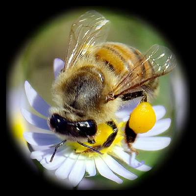 There are similarities between humans and bees. Psychologists confirms that human in nature prefer natural substances for healing diseases, so we find human by nature accept honey and other natural substances more than the chemical ones..
USA 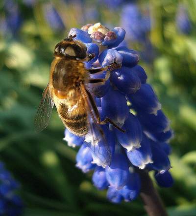 Honey has ‘information’ that passes from the bee to the honey during the production process. This information is found in the flowers’ nectar. Inside the belly of the bee, this information is interacted, modified, and increased in effectiveness so it becomes ready to use, and this is the secret of healing with honey. Allah the Almighty provided each bee with a special programs in its brain cells, which means that it performs something that has been already planned for it, and this is what is expressed in the Holey Qur’an: “And your Lord inspired the bees.”. This is a way planned earlier for the bee, it is an inspiration from Allah the Almighty in a method that we human can not understand!
Miracle in the Qur’an and SunnahSurat
http://www.youtube.com/watch?v=VtHTl1muZ40
http://www.youtube.com/watch?v=r1KTNG8oVLc
THIS IS ONE EXAMPLE AND SOME BENEFIT THAT YOU CAN LEARN TO PROVE AND DISCOVER ANYTHING.
LET'S BECOME A MICROBIOLOGIST FOR OUR DEVELOPMENT IN OUR COUNTRY AND LET'S EXPLORE IT! :)
-EVA NATASHA-
According to a recently published study by a team of prominent researchers from the College of Medicine & Health Sciences at United Arab Emirates University (UAEU), honey has dramatically beneficial effects in cancer treatment. The ground breaking discovery provides strong scientific evidence that manuka honey, obtained from nectar collected by honey bees from the New Zealand manuka tree, can effectively inhibit the growth of a variety of cancer cell types, including breast, skin and colon cancer. Additionally, the study highlights a new and potentially very exciting property of manuka honey in reducing the toxic side effects associated with chemotherapy treatment in cancer patients.
The study was led by Dr. Basel al-Ramadi, Professor and Chair of the Department of Medical Microbiology and Immunology, College of Medicine and Health Sciences and the study, entitled “Intravenous administration of Manuka honey inhibits tumor growth and improves host survival when used in combination with chemotherapy in a melanoma mouse model”, was published in the renowned scientific journal PLOS ONE.
Dr. Al-Ramadi explained, “Manuka honey has been recognized for its anti-bacterial and wound healing properties for many years. However, the potential effect of manuka on cancer cells has not been investigated in detail. In this study, the team of researchers used three different cancer cell lines of human or murine origin and demonstrated that the addition of exceedingly small amounts of manuka honey, as little as 1.0 %, can stop the growth of cancer cells. The researchers then carried out an extensive series of experiments to uncover the molecular basis of manuka’s anti-cancer activity. Our findings provided conclusive evidence that manuka acts directly by inducing apoptosis, or programmed cell death, in cancer cells. Apoptosis is a physiological process that all multicellular organisms use to balance their need for new cell generation with the elimination of old unwanted ones. This process is tightly regulated so that, in adult tissues, cell death exactly balances cell division. If this were not the case, excessive apoptosis causes tissue atrophy, whereas insufficient apoptosis would lead to uncontrolled cell proliferation, such as in cancer. Hence, manuka induces the death of cancer cells through the same physiological process used by our body to maintain normal cell number.”
In the course of their investigation, which began more than 5 years ago, the researchers also used an animal tumor model to demonstrate the potential effect of manuka honey in vivo. The results of these experiments confirmed the usefulness of using manuka as an anti-cancer agent. Remarkably, however, when the researchers used manuka honey in conjunction with a standard chemotherapeutic agent, they noticed that the combined treatment resulted in a significant improvement in overall animal survival. Based on these findings, the investigators concluded that the combination treatment with manuka plus anti-cancer drugs maintains effective anti-neoplastic therapy while simultaneously reducing the toxic side-effects of chemotherapeutic agents.
"This is a very exciting area of research and we are optimistic about what these new developments may mean in terms of potential new treatments for certain types of cancer,” added Dr. Al-Ramadi.
Dr. Ali Rashid Al Noaimi, UAEU Vice Chancellor, commended the team of researchers and said, “Following the vision of H.H. Sheikh Nahayan Mabarak Al Nahayan, Minister of Higher Education and Scientific Research, Chancellor of UAEU to be a leading Research Intensive University of International Stature, our world-class faculty continue to demonstrate the abilities and strengths of this University in addressing the significant challenges faced world-wide and continue to contribute significantly in many areas in particular medical advancements. The very fact that this research has been published in such a highly regarded publication is testament to its importance, and testament to the brilliant work that Dr. Al-Ramadi and his colleagues have done. We are very proud of their achievement.” The group of investigators also includes Dr Hakam El-Taji, formerly a senior consultant of surgery in Tawam Hospital, Dr Maria Fernandez-Cabezudo (Department of Biochemistry) and Dr Fawaz Torab (Department of Surgery), UAEU College of Medicine and Health Sciences.
The experimental findings reported in the recent publication are expected to stimulate further investigations on the use of manuka honey in patients that would hopefully usher in new modalities for cancer treatment in humans.
Dr. Molan found that all kinds of honey have strong antibiotics; he said that there is no single substance in the world that has antiseptic properties like honey, because the bees produce a stuff called Hydrogen Peroxide through special enzymes that is known for its antiseptic properties.
After 20 years of experiments, this researcher proved that honey has high healing power that can be used for treating prolong constipation without any side effects. He says that the medical tools he carries in his treatment case are just honey and bandages! Dr. Molan treats so many diseases just with honey! And he says: “honey has a wonderful effect when treating burns and suppuration, it can be applied directly on burns to repair the skin and kill the bad bacteria, it removes the burn marks and you can see the burned part without marks or scars”
There are similarities between humans and bees. Psychologists confirms that human in nature prefer natural substances for healing diseases, so we find human by nature accept honey and other natural substances more than the chemical ones..
After long experiments, Cancer specialist – Dr. Glenys Round- discovered something new about honey! He discovered the amazing effect of honey in treating cancer. He said that he used honey in treating skin cancer because honey penetrates through the skin and treats the cancer; something other medications failed to do.
He also confirmed that all the drugs failed to cure ulcers but finally ulcer is cured when honey was used. All the patients who were treated by honey were so happy during the treatment because they had no side effects and no pain.
In the USA
Experts confirm that 6 Billion Dollar is spent per year on treating injuries and burns, and a big percentage of this money can be saved if honey is used instead, so honey can save money too.
Researchers found that honey had a healing power for treating stomach ulcer and throat infection. They found that germs usually stick together in a way to assure their existence and concentration, the scientific research proved that honey aparts, disintegrates, and weakens these germs defences so the body can kill them. Lately, the scientists discovered stuff in honey that stops oxidation and therefore honey helps treating Cholesterol
Honey has ‘information’ that passes from the bee to the honey during the production process. This information is found in the flowers’ nectar. Inside the belly of the bee, this information is interacted, modified, and increased in effectiveness so it becomes ready to use, and this is the secret of healing with honey. Allah the Almighty provided each bee with a special programs in its brain cells, which means that it performs something that has been already planned for it, and this is what is expressed in the Holey Qur’an: “And your Lord inspired the bees.”. This is a way planned earlier for the bee, it is an inspiration from Allah the Almighty in a method that we human can not understand!
Scientists wonder about the hidden healing power found in honey which can treat incurable diseases, and they ask: how does healing happen? What does honey do inside the cells of our bodies that lead to a sudden stop of cancer, cease the growth of so many bacteria in the body and activate the immunity system and make it more efficient? What does happen? No one knows
Miracle in the Qur’an and Sunnah
It is narrated in the Sahihan that “A man came to the Prophet peace be upon him and said, ‘my brother is complaining about his stomach’. The Prophet peace be upon him said, ‘give him some honey.’ The man went and came back later, saying, ‘I have given him some honey, but it did not help.’ He repeated this three times, all the while the Prophet peace be upon him continued saying to him, ‘give him some honey.’ In the fourth time he came, the Prophet peace be upon him said, ‘give him some honey.’ And the man said ‘I have given him some honey, but it did not help.’ Then the Prophet peace be upon him said, ‘Allah has said the truth while your brother’s stomach has lied.’” The man gave his brother some honey and he was healed.
The Prophet’s words above indicate the importance of using honey in treating digestion disorders.
The stomach complain that came in the Hadeeth above is the closest translation for the original Arabic word ‘istitlaq’, which is known today to be diarrhea. Studies showed that honey kills all types of germs especially the ones exist in the digestion system. As a result, honey is considered effective in treating Diarrhea and stomach ulcer in a very short time. However, honey has a dual effect, it also treats constipation, it regulates the motion of the colon and influences the colon with the information that Allah the Almighty has designed it with.
Finally, we want to mention a hadeeth [narrated by Al-Bukhari] stated that the Prophet peace be upon him said, “There is cure in three substances, a drink of honey, a slash with a knife used for cupping and cauterizing by fire. I forbid by nation from cauterizing by fire.”
Another hadeeth [narrated by Ibn Majah and others] stated that the Prophet peace be upon him said, “Make use of the two cures: honey and the Qur’an”
We recall Allah the Almighty says: “There comes forth from their bellies, a drink of varying colour wherein is healing for men. Verily, in this is indeed a sign for people who think. (An-Nahl: 69).
I strongly suggest we start using honey and the Qur’an for healing, something the Prophet peace be upon him advised us to do, then we can follow the path to healing. From experience, when we recite Surat
http://www.youtube.com/watch?v=r1KTNG8oVLc
-EVA NATASHA-